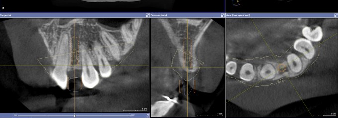
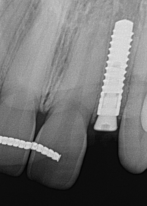
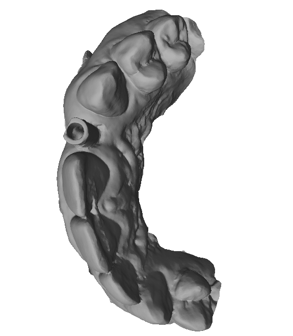
Would you do this implant as planned and thinking ahead...

Here is the implant plan. A very tight spot and high "pucker" factor. Would you place the implant as planned and if so. If not, how would you change the plan? Or... do you think the space is too tight and would not place the implant at all... She has already been through Ortho...
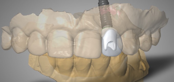
If you would place the implant, what are some of the considerations you have to be thinking about moving forward about the restoration phase?


High pucker factor - Yes! I always go with my first reaction and when I looked at your screen shot, I thought "Guided could get this done." I would explain the risk to patient and let her decide. My mouth, your skillZZZZ - I would not be worried. Good Luck Skrammy.
*Side note - looks like a 3.3 Straumann, any chance you would change to different implant and go to 3.0?
Krafty
Hi Mike, Not the most experienced but I will venture an opinion. I'd would like to see photos as well, but based on what you show I can see an fully guided surgery placing an implant accurately in the planned position (assuming the sleeve shown fits without interference). I do not like how close the implant is to the adjacent teeth and consider it a high risk for complications. I have done a number of these cases successfully. I prefer an implant diameter similar to what you show for ease of prosthetics, but I'd prefer a more narrow implant from the surgical viewpoint to decrease risks. In my hands those style implants are far more challenging to restore. Nice case to stimulate thinking though

I agree very tight space there - might be a nice case for a tapered implant like the 3.0 biohorizon implant - clinical photos would be great tool to assess as well, sure your got 'em must be holding back for some reason.
Thinking ahead for the restorative phase it looks like you will have a lot of room to make up from the implant platform to the restoration - this running room will be nice to create emergence but will you have to profile the bone given the depth of the implant placement. I am sure you will reveal some wonderful result or great teaching point and I look forward to learning from this case.

From a purely surgical approach, I would have no problem with this implant being placed. You are a safe distance from the buccal plate in the coronal 1/3, so I wouldn't worry about losing the buccal wall. You may give some thought to grafting the buccal concavity, but only if needed cosmetically. As planned, it looks like the sleeve on your guide will intersect with the canine. I would need some intra oral photos of the space and contra lateral tooth to evaluate for the restoration.

Ok... I agree. The implant was close, but doable. I talked to the patient about possible risks being a little closer to ideal to the adjacent teeth and we decided to move forward...
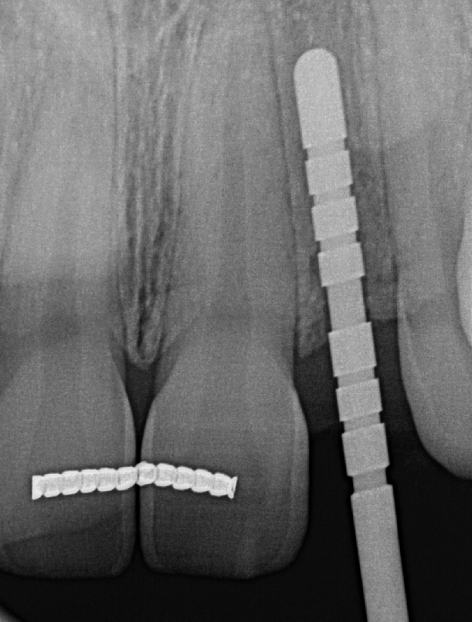

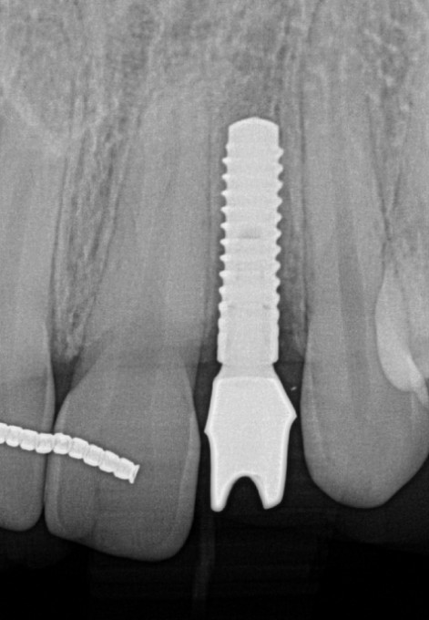
Final image is a little deceiving. The angulation makes it a bit closer to central. It may be a little closer apically to the central, but that has proven not to be super critical in the research. It was placed with CG2
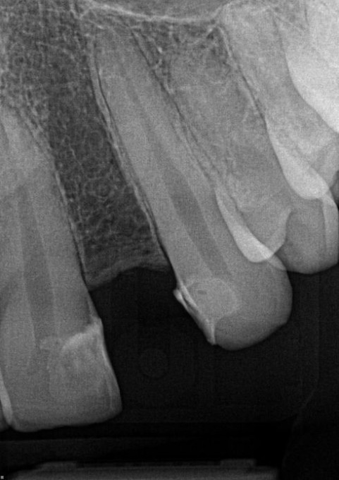
She was wearing a flipper, so I placed a very small healing cap that went subgingival so i did not have to adjust flipper. Basically am burying it for 4-5 months.
Now... how do you restore. This is the hard part. It's very tight. Will the Sirona Tibase work?

Mike,
The restorative phase is challenging(with your skills -no problem). Are you going to do a little enamoplasty on the mesial of the cuspid to gain a little room?
Yes it's doable. I would look into other implant sizes. A tapered 3.0 would be ideal.
Here's a similar case to yours, Mike. My go-to implants are Straumann Bone Levels. However with this post ortho case, I didn't feel comfortable with the 3.3 diameter. Therefore, I chose a 3.0 BSB Max with the Nobel Active NP connection. It can still be restored with a tibase, so restorative work flow with CEREC should be fairly predictable.
Mike,
I'm thinking ti base is going to be too wide at the implant interface.We have seen this on the boards before. I think Farhad posted something of this nature in the past. I would have a working model with lab analogue with soft tissue. You can then prep on the model for a temp with a temp abutment. When the implant is ready to be uncovered, the temp can be inserted. Sometimes traditional impression with an impression post is the correct choice. That being said, It doesn't hurt to try a digital restorative pathway. I'm sure you will nail it as always.
Paul

This case is part of Mike's lecture at the upcoming implant seminar in a couple of weeks so I will not comment on it too much here yet. His lecture will complement the challenging lateral incisor case we will be doing as the live surgical demonstration.
I personally would not have a problem placing a 3.3mm implant here as planned but Straumann just released a 2.9mm implant in Europe, which will be great for these situations (should be available here in the U.S. in the fall).
Farhad
My lecture at the Implant Seminar with be "The tale of 7 laterals". I'm going to go through 7 different clinical scenarios that I treated a little bit differently with laterals. It's a hard tooth to treat with implants. I will include two that did not work at all as well and why "i think" that was.
As far as this case, I will show you what I did for sure. Just give me a minute.

The main difficulty of this case is that there is very minimal bone around the coronal portion of the implant. Any stress on the tissue and especially bone will make an already risky case especially prone to complications. If you look at the radiography, there is absolutely no way to profile the bone here. Even if you could profile the bone, the shoulder of the titanium base (whether it's Sirona's, BSB's, or even Straumann's) is just too wide at that depth. Not only would it greatly increase the risk of biologic complications, but it also would be almost impossible to restore.
Farhad has shown a case similar to this in the past. Since we know we absolutely cannot use one of the above listed titanium bases, we have to think of different options... One option would be to simply take an implant level impression and send it to the lab for a traditional custom abutment. While this will work... come on man :) We are not going to do that!
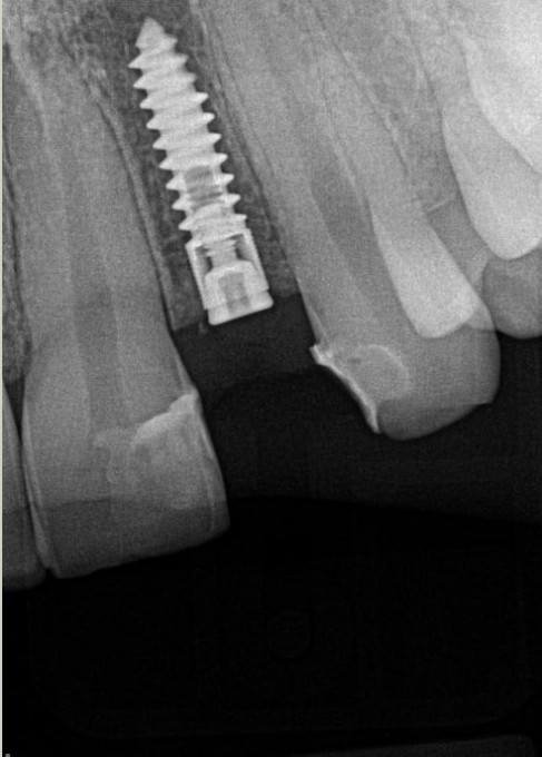
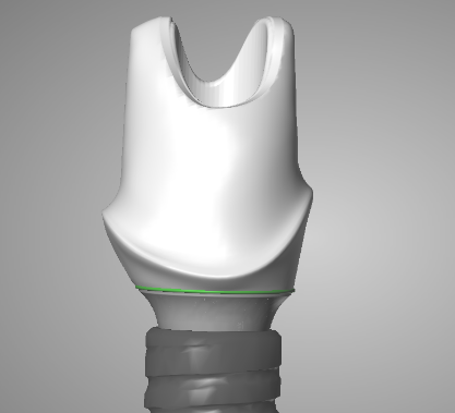
The plan was to digitally create a custom zirconia hybrid abutment using a Straumann Variobase with a much higher collar. In order to do this, you will need to do a couple things that will be new to most...
- You need to order a Scan Body from Straumann (not use a Sirona Scan Body)
- You will need to find a laboratory that has BOTH inLab 15 AND either Dental Wings or 3 shape software. There are many out there that have both... i don't want to specifically promote one lab here.
- Scan the Straumann Scan Body intraoral as normal. Upper, Lower, Gingivamask, and Buccal Bite
- Send to your Lab via CEREC Connect. They can import into inLab 15 and convert the file to .stl. This .stl can be imported into another software that communicates with Straumann's official milling center in Arlington Texas.
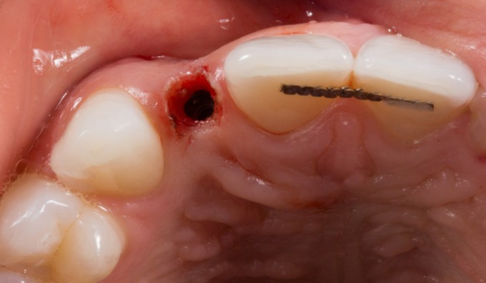
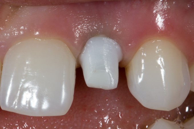
Here are the clinical pictures:
Uncovered the implant:
Placed the Straumann ScanBody:

.stl Conversion of file:
Design of custom abutment:
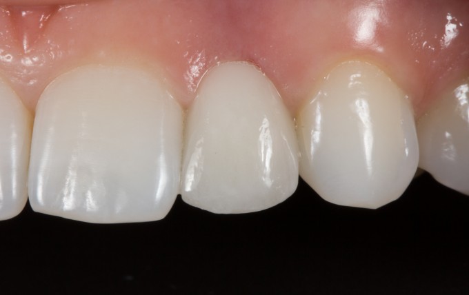
Abutment seated in mouth:
Final Restoration:
I show this case to show some of the limitations of what we currently can do with CEREC. This will change very shortly as both Sirona and other companies have recognized the weakness of the current titanium base system on narrow diameter implant. I think you will see Straumann's new tibases in the software by the end of the year and Conelog also has some solutions as well... with more to come i'm sure.
However, we also have to recognize when we need to send something to the lab to clinically get the right result. Fortunately for us, there is a digital way to do this... because I don't remember how to take an impression :)
Thank you Mike for the case! I think you are the first one show the case with different software and CEREC. Everyone who placed the implants knows how tough to restore narrow implants. I think Sirona has to give us more flexibilty restoring cases like yours. Gregory

On 5/5/2016 at 9:02 am, Gregory Mark said... Thank you Mike for the case! I think you are the first one show the case with different software and CEREC. Everyone who placed the implants knows how tough to restore narrow implants. I think Sirona has to give us more flexibilty restoring cases like yours. Gregory
They will... Sirona now has a firm understanding of the need.
On 5/5/2016 at 8:52 am, Mike Skramstad (Faculty) said...I show this case to show some of the limitations of what we currently can do with CEREC. This will change very shortly as both Sirona and other companies have recognized the weakness of the current titanium base system on narrow diameter implant. I think you will see Straumann's new tibases in the software by the end of the year and Conelog also has some solutions as well... with more to come i'm sure.
That's awesome to hear. Would love having flexibility with additional tibase designs
What's an impression?However, we also have to recognize when we need to send something to the lab to clinically get the right result. Fortunately for us, there is a digital way to do this... because I don't remember how to take an impression :)


On 5/5/2016 at 9:57 am, Alex Botvinnik said... This is really beautiful, Mike.
Can you think of a similar workflow with a Nobel 3.0?
Similar workflow as Mike has outlined but:
1.) Instead of the Straumann scanbody use a Nobel 3.0 scanbody, either from Nobel directly or a 3rd party company like NT Trading or Dominion milling (I don't think Glidewell has a scanbody for the Nobel 3.0).
2.) Find a lab that has inLab 15 with STL export license and has a restorative design software with the implant library corresponding to the scanbody you used (Procera, Dental Wings, ExoCad, 3Shape).
Farhad

I do not think a BLT allows you any more restorative wiggle room here Chuck.
You have to be kidding Mike right ;)
Thought never crossed my mind

can u tell me about the abutment..does it come in two pieces or do they deliver it as one piece,, and the name of that base or abutment
On 5/5/2016 at 2:58 pm, marc kaufman said...can u tell me about the abutment..does it come in two pieces or do they deliver it as one piece,, and the name of that base or abutment
Custom milled zirconia abutment (designed in Dental Wings and milled at Straumann CARES milling facility) and Straumann Variobase for CARES (NC in this case). It will be delivered in 2 pieces and you have to bond together as usual.
Farhad

Great case Mike! Impressive in regard to the surgical and restorative challenges. I'm assuming you seated the abutment and then imaged that and then made the crown that day?
Yes, I reimaged and made the crown. There is ways to split the case in other software and have them make the crown off the abutment design, but I like the control.

Mike,
The healing abutment xray shows great crestal bone, but the abutment xray appears to show some crestal bone loss. Is this clinically the case? If so, to what do you attribute that to?
Rich, there might be just a little, but not much. It's the xray. I have a 3D post op as well and it looks fine.
Mike, So cool to see a case like this, testing the limit of space between implant and neighboring roots. It's been over 3 years. Can you please share an update on this patient? Radiograph, full smile and retracted anterior photos would be terrific to see. Thank You!