CBCT Shines a Light on Every Day Diagnosing and Treatment Planning

Dan Butterman, DDS
One of the most valuable tools we have available for diagnosing and treatment planning is CBCT imaging. It can be very difficult to diagnose what you can’t see, and it is impossible to treat what you don’t diagnose. CBCT imaging shines a light on otherwise difficult diagnoses and asymptomatic pathology.
In my practice, 3D imaging is not only necessary for implant planning, but also for routine diagnoses. My full series of radiographs consists of bitewings and a scan with the Axeos 3D CBCT. The scan is instrumental for a comprehensive patient exam. The Axeos gives me the complete diagnostic package, including larger fields of view and low dose scans.
The larger field of view enables me to visualize the patient’s airway, maxillary sinus, and TMJ. The low dose scan, which has a similar radiation dose to a 2D image, gives me an accurate post op analysis of treatment such as implant placement and endodontic therapy. Implants and root canals can fail for a variety of reasons. In today’s litigious society, it’s a good idea to have a 3D post op confirmation of proper implant placement and endodontic obturation.
Whether you’re contemplating incorporating the Axeos or another CBCT in your practice, here are a few examples of how the technology will pay off for you and your patients on a daily basis:
- The patient presented with gross caries on tooth #3. While the periapical radiograph shows no definitive evidence of periapical pathology, the CBCT tells a different story: there is a lesion associated with tooth #3 infiltrating the maxillary sinus.
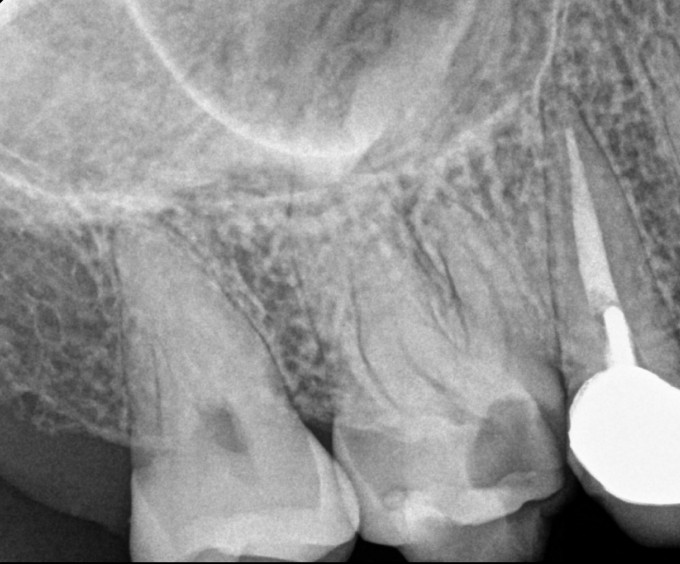
Periapical radiograph of gross caries tooth #3
CBCT slice visualizing palatal root lesion infiltrating the maxillary sinus
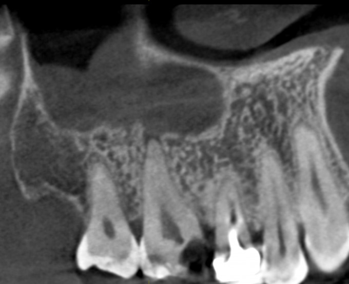
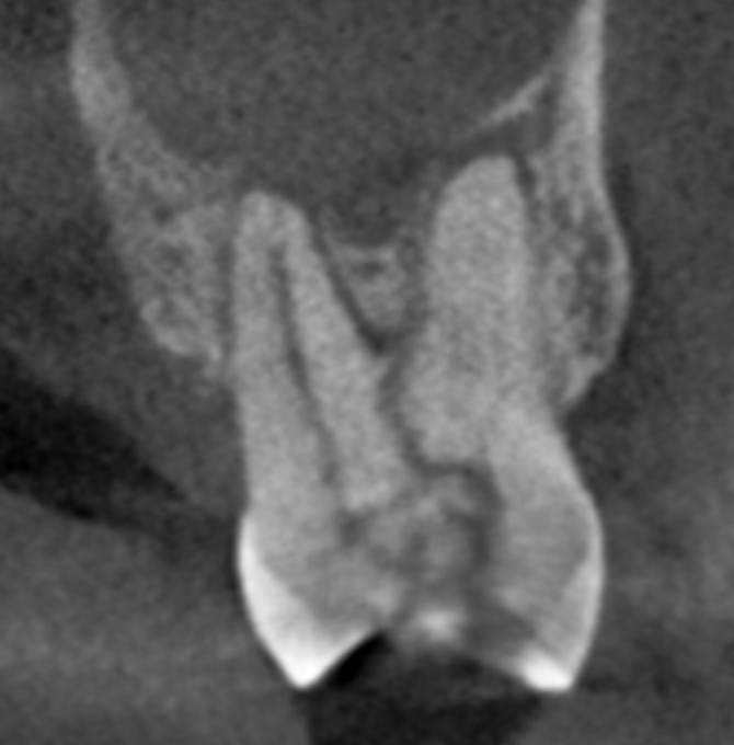
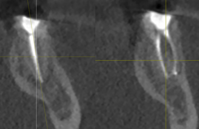
- The patient presented with a fracture line through the central fossa of tooth #2. The periapical radiograph is inconclusive, but the CBCT demonstrates the extent of the fracture and the fact that tooth #2 is non-restorable. (figs 2 A-D)
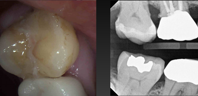
Intra oral picture and bitewing of fractured tooth #2
2D Periapical radiograph of tooth #2
Cross sectional view of CBCT demonstrating extent of vertical fracture and sinus lesion
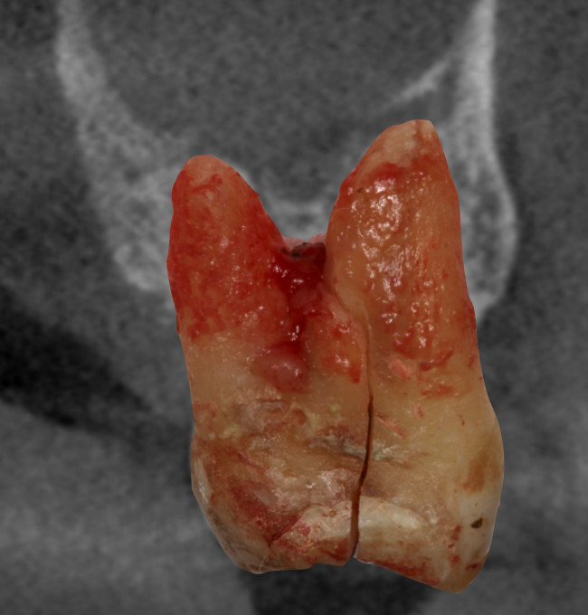
Extracted tooth #2 with vertical fracture
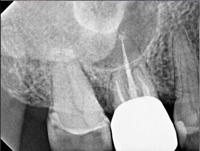
- The patient presented for a second opinion of tooth #18. Based on the periapical radiograph, which shows a radiolucency on the distal root, the patient was scheduled for an endodontic re-treatment of tooth #18. The CBCT shows that the presumed radiolucency is actually the mandibular canal superimposed on the distal root. The CBCT also shows that the true pathology is a vertical fracture and that tooth #18 is non-restorable.
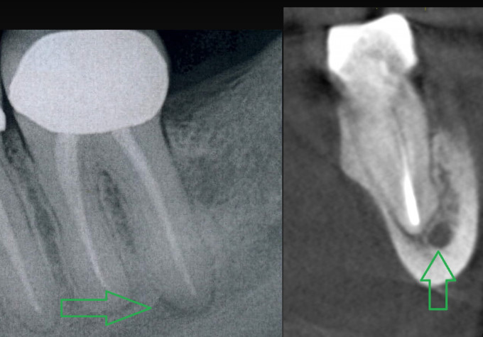
Periapical radiograph of tooth #18 showing increased radiolucency at distal root. Cross sectional CBCT view of tooth #18 showing normal distal root PDL with the position of the mandibular canal
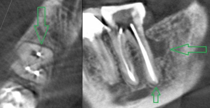
Axial and Tangential CBCT view showing fracture and mid root lesion
- Low dose scan for evaluating post endodontic obturation.
Cross Sectional Low Dose CBCT view confirming 3D endodontic obturation of all canals
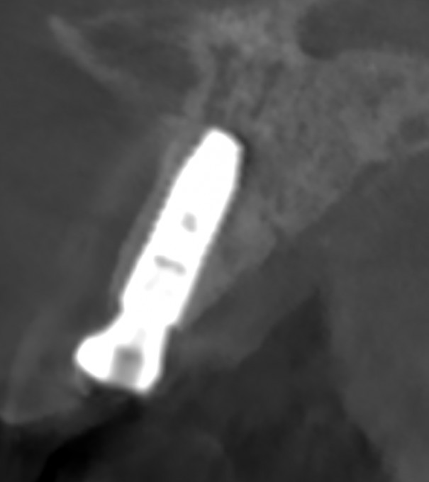
- Low dose scan to confirm implant position after guided surgery.

Cross Sectional Low Dose CBCT View confirming proper implant placement based on plan
- Low Dose scan to evaluate post op implant placement in the anterior.
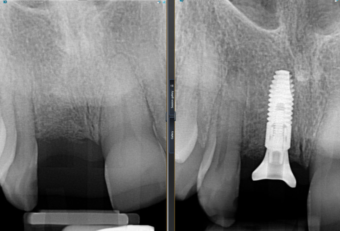
2D periapical pre-op and post- op implant placement at site #8

Low Dose Axeos scan reconstructed 3D view
Cross Sectional Low Dose CBCT view confirming post op implant placement in relation to the buccal plate
- Routine scan demonstrating possible airway issues in a patient that reports poor sleep.
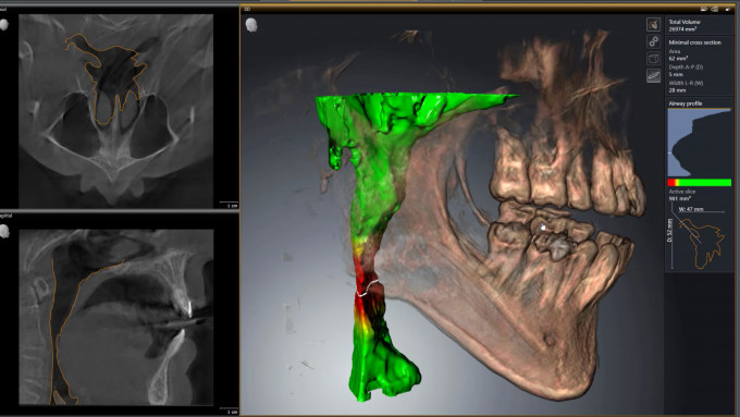
Airway analysis view in SiCat 2.0 Airway module
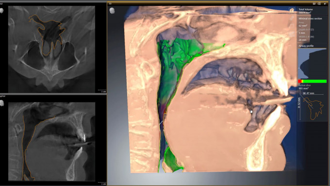
Airway analysis view with soft tissue in SiCat 2.0 Airway module
These cases are every day examples of the benefits of taking a routine CBCT scan in place of a full series of periapical films. We no longer have to “watch” and wait for a tooth with vague symptoms to become an issue. With CBCT we can accurately evaluate the tooth before it blows up. A CBCT scan also gives us a better understanding of tooth’s prognosis before treatment is initiated. Upon completion of treatment, a low dose scan gives us confirmation that endodontic therapy and implant placement were performed successfully.


From Day one I put my Axeos to use in the low dose and high resolution rendering. Could identify a hidden abscess!

It is surprising how often we find pathology in a 3D image that just wasn't possible to see in a 2D image.