CONE BEAM Video
How to Read a CBCT Scan – Step-by-Step Volume Review
Dr. Steven Hernandez
CDOCS Faculty

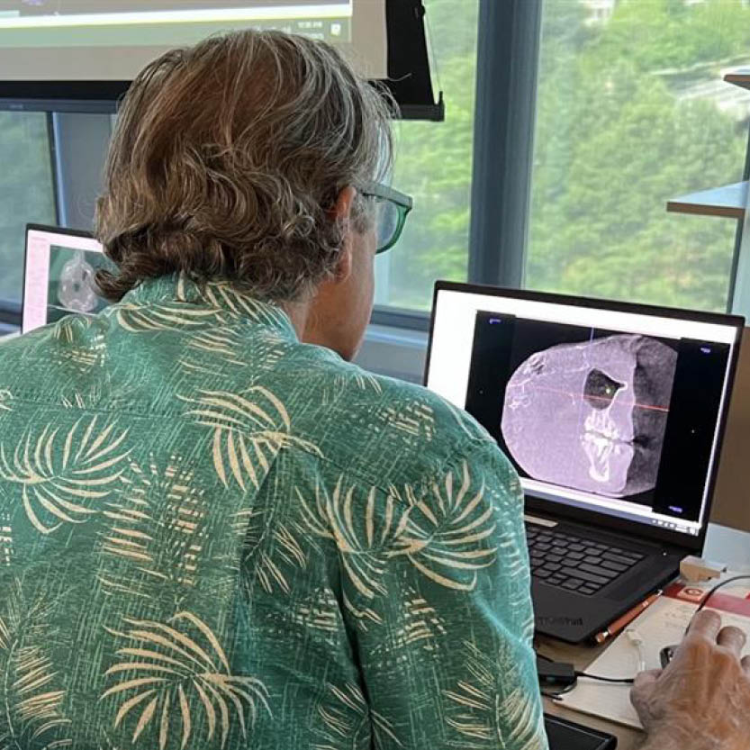
This tutorial teaches a systematic three-pass volume review method — coronal, axial, and sagittal — designed to ensure no pathology is missed during a CBCT examination. Dr. Hernandez also demonstrates how to compare two scans side by side to track changes in bone levels and lesion size over time.
- Open the multiplanar view and remove the crosshairs to reduce visual clutter.
- Scroll coronally, front to back, comparing the left and right sides for asymmetry, the most useful diagnostic signal on a scan.
- Scroll axially, top to bottom, noting anything unusual in the sinuses, airway, or roots.
- Switch to the sagittal view, left to right. This angle most closely resembles a periapical film and is the easiest for spotting periapical lesions.
- If something looks different from a prior visit, use the Compare tool to pull up the patient’s previous scan side by side, desynchronize the two views to align them, then resynchronize to confirm what changed.


Related CDOCS Hands-On Workshops
Implementing Cone Beam for Practical Utilization (CB110)
Get comfortable with cone beam technology through hands-on training in CBCT hardware operation, full volume review, and Sidexis and Sicat software suites, including introductions to CAD/CAM integration, airway analysis, and implant and endodontic applications to unlock your system’s full potential.

Maximizing Diagnostic Efficiency with Cone Beam (CB210)
Enhance your ability to systematically review CBCT volumes, identify and categorize radiographic abnormalities, and effectively collaborate with maxillofacial radiologists.