CONE BEAM Article
Maximizing CBCT Utility with AI Integration
Dr. Steven Hernandez
CDOCS Faculty

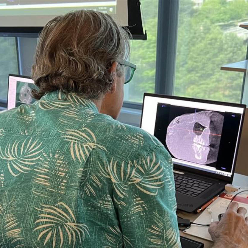
For dental professionals who have integrated Cone Beam Computed Tomography (CBCT) into their practices, the technology quickly shifts from luxury to a daily necessity. Over the 13 years I have utilized 3D imaging, it has become the cornerstone of my clinical workflow. While many initially invest in CBCT for the precision required in implantology, its utility extends far deeper into the realms of endodontics, oral surgery, and the critical evaluation of patient airways for Obstructive Sleep Apnea (OSA).
However, the challenge has never been the acquisition of the data, but rather the efficiency of its interpretation. This is where the digital landscape is shifting. DS Core is evolving at a rapid pace, transitioning from a simple storage and viewing platform into a robust diagnostic ally.
From Static Viewing to Dynamic Analysis
In the early iterations of DS Core, the functionality was largely restricted. We were able to scroll through a volume, but the deep diagnostic tools remained tethered to desktop-bound software like Sidexis. That barrier is now dissolving.
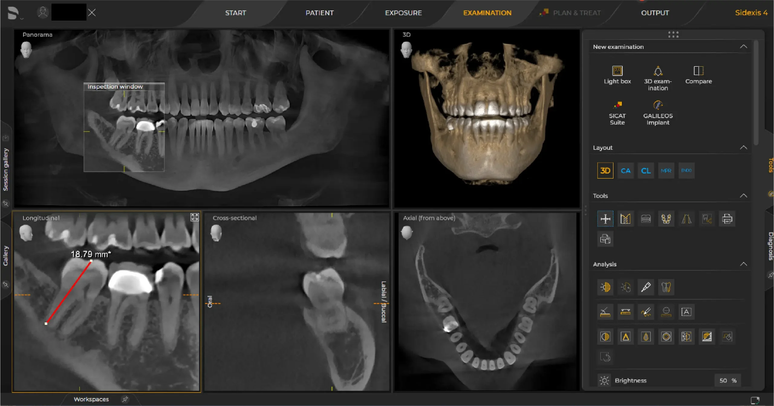
The Canvas feature within DS Core has revolutionized how we interact with 3D data. Beyond simple viewing, we now have the ability to perform precise measurements directly within the cloud-based volume, just like in Sidexis.
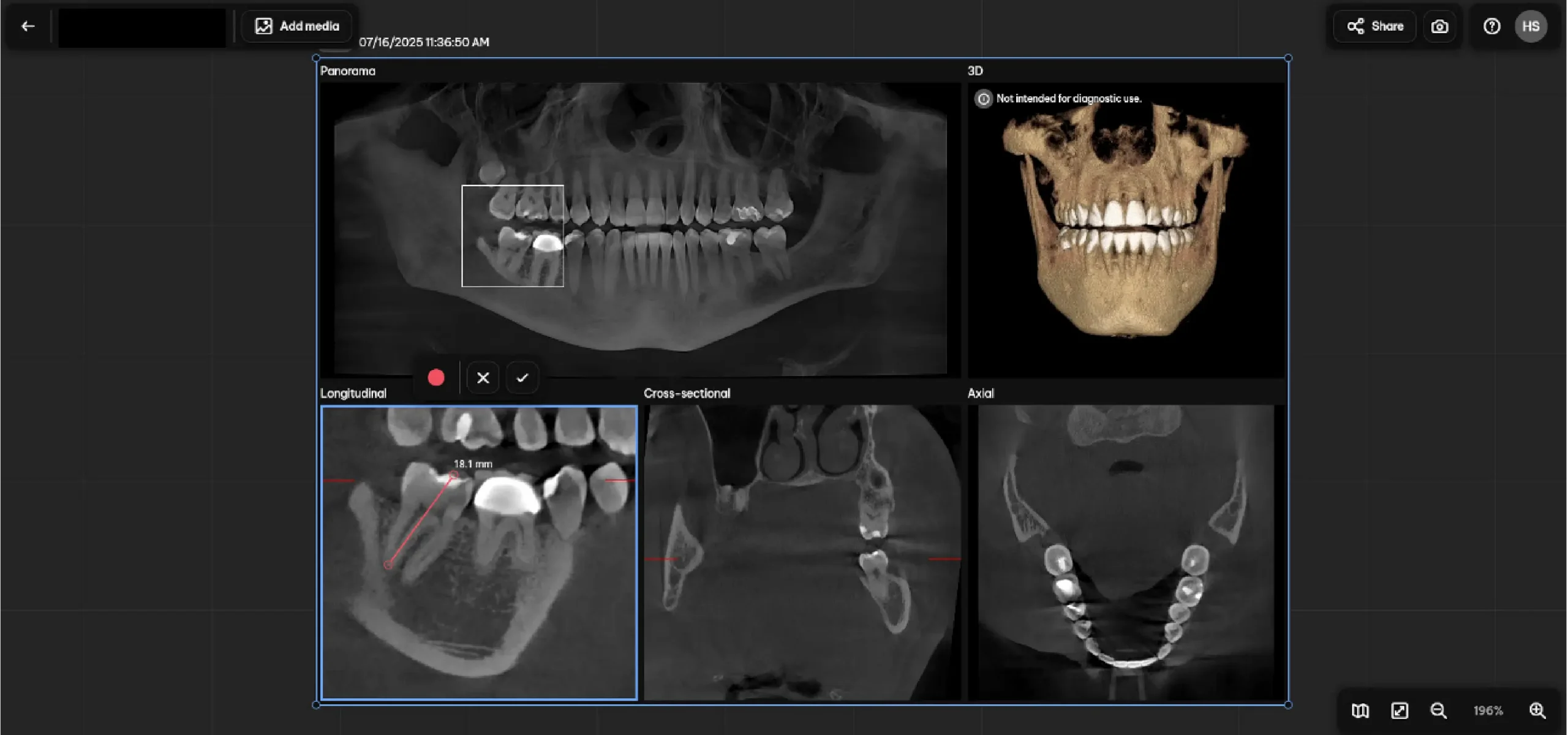
Furthermore, the AI Smart View capability allows us to distill complex 3D data into a format that is far more patient friendly. This is a crucial step in patient education; by presenting a clear, simplified visual, we bridge the gap between clinical diagnosis and patient acceptance.
The New Frontier: AI-Driven Pathology Detection
Furthermore, the AI Smart View capability allows us to distill complex 3D data into a format that is far more patient friendly. This is a crucial step in patient education; by presenting a clear, simplified visual, we bridge the gap between clinical diagnosis and patient acceptance.
Navigating the New Workflow
To integrate this into your daily routine, the workflow is designed to be intuitive:
1. Accessing the Volume: Within DS Core, select the specific patient volume you wish to analyze.
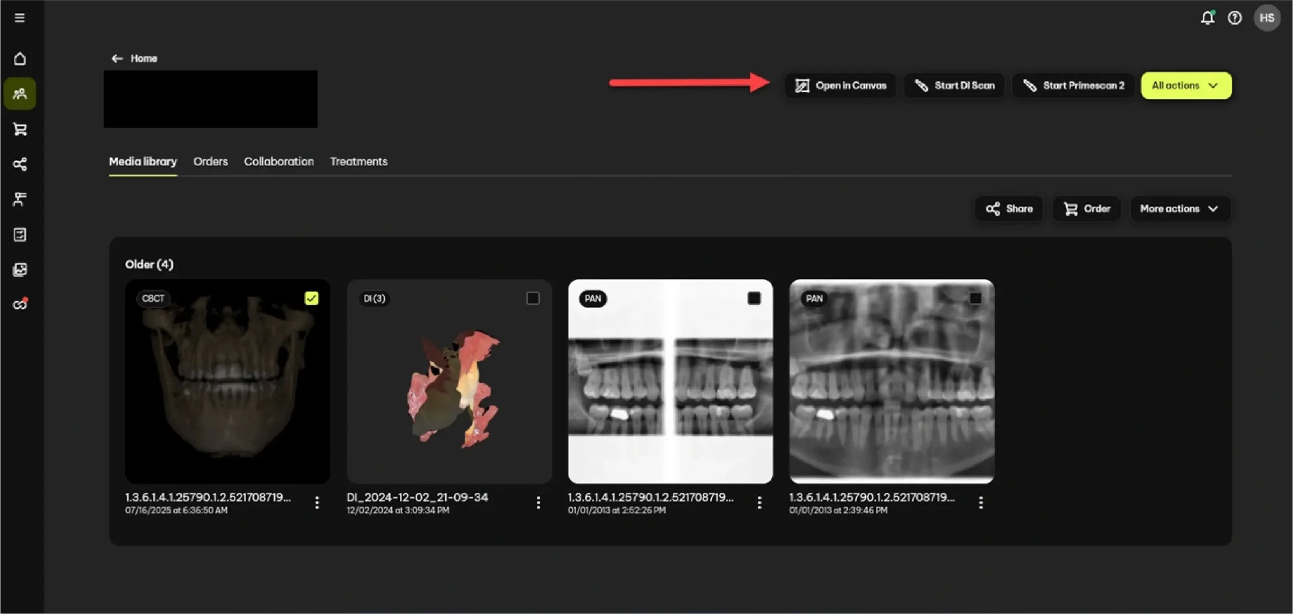
2. Launching Canvas: When the volume opens in Canvas, it will mirror the default 3D layout familiar to Sidexis users, ensuring a minimal learning curve.
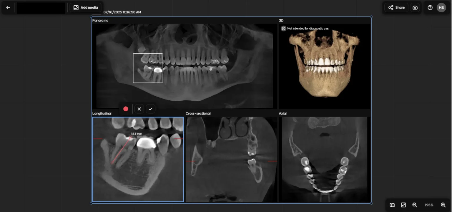
3. Activation Time: Once the volume is open, allow approximately one to two minutes for the AI icon to become active. This brief period allows the software to process the complex data in the background.
4. Automated Detection: Selecting the AI icon triggers the software to automatically scan the volume and highlight potential PARL’s for your clinical review.
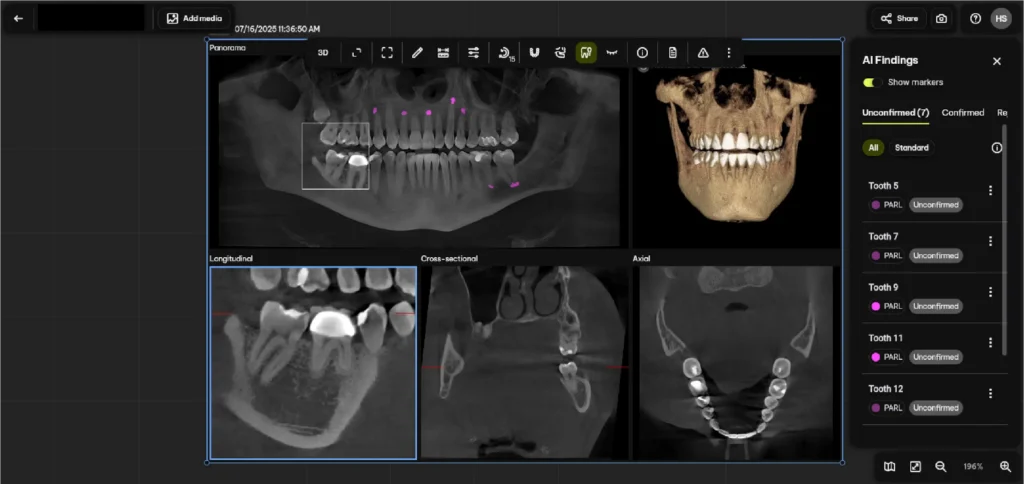
Precision through Sensitivity Settings
The software offers a nuanced approach to detection with two distinct settings: All and Standard.
- All: This setting provides a highly sensitive reading of the volume. It is designed to catch even the most subtle changes in bone density, ensuring that no potential pathology goes unnoticed.
- Standard: This results in a more moderate sensitivity level, filtering out minor irregularities to focus on more pronounced clinical findings.
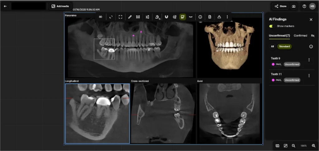
To review a flagged area, simply double-click the notification. The software will automatically orient the view so the area in question is centered. From there, you can utilize a slider button to toggle the AI overlay on and off, allowing you to compare the raw data with the AI’s findings.



Conclusion
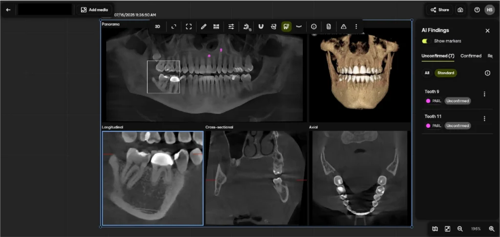
It is important to remember that AI is a tool for augmentation, not replacement. In a recent case, the software flagged an area for review; however, upon closer inspection using the toggle tool, I determined the tooth was healthy and no pathology existed. The platform allows for this clinical autonomy, providing three dots next to the finding where you can either confirm or reject the AI’s suggestion.
This update is a monumental step for the dental community, particularly for those hesitant to adopt 3D imaging. By providing an automated starting point for volume interpretation, DS Core is lowering the barrier to entry and helping doctors become more proficient in 3D radiology. While no software is perfect, this AI integration represents a sophisticated first step in making comprehensive 3D diagnostics more accessible, accurate, and indispensable than ever before.
Related CDOCS Hands-On Workshops
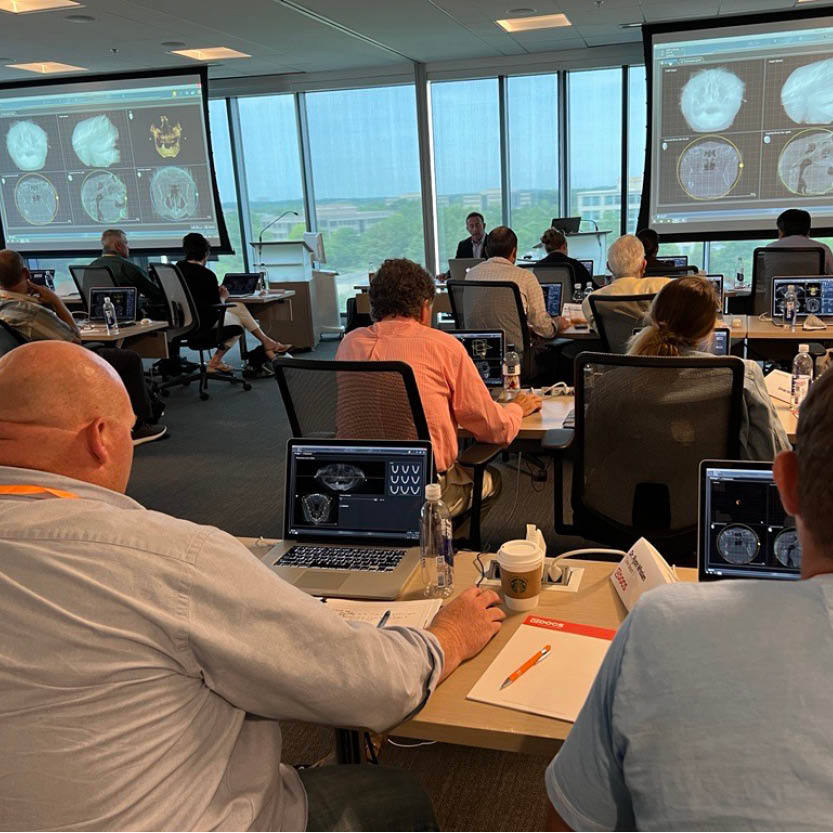
Implementing Cone Beam for Practical Utilization (CB110)
Get comfortable with cone beam technology through hands-on training in CBCT hardware operation, full volume review, and Sidexis and Sicat software suites, including introductions to CAD/CAM integration, airway analysis, and implant and endodontic applications to unlock your system’s full potential.
Maximizing Diagnostic Efficiency with Cone Beam (CB210)
Enhance your ability to systematically review CBCT volumes, identify and categorize radiographic abnormalities, and effectively collaborate with maxillofacial radiologists.