 Rich Rosenblatt, D.M.D.
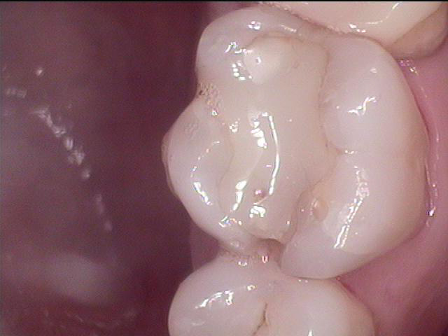
 I recently purchased the Sirona XG3D 2D pan/ 3D cone beam. It is quickly changing my practice and my ability to diagnose my patients. A patient came in yesterday. I took BW's during his checkup. Upon exam we saw this tooth with a chipped interproximal composite that had cracks on the distal. I was debating b/t an onlay and crown. He had no discomfort on tooth previously and he didn't even notice that it was chipped. I took a PA for insurance purposes.
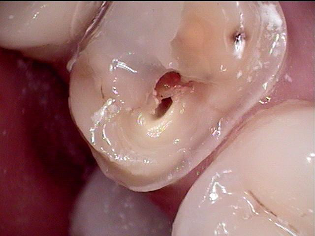
This is the pre op PA with my new schick 33's. It looks like maybe some decay under the composite.
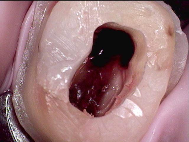
I show him this photo and tell him that I'm going to have to do endo. I put composite into that space, prep it to ideal and image to make crown while doing endo. I then open a new window and image the prep as a pre op so I can make a biocopy of the core and just bond that right in when the endo is completed. After both images are done, I create an endo access. The m and d canals are necrotic and the palatal canal looks huge and it is bleeding like crazy. I could not get it to stop. I inject some septocaine into what I thought was the canal and get some hemostasis but not total. I take a pic and this is what I see. (all these pics are just with my IOC since I was not planning on documenting this one):
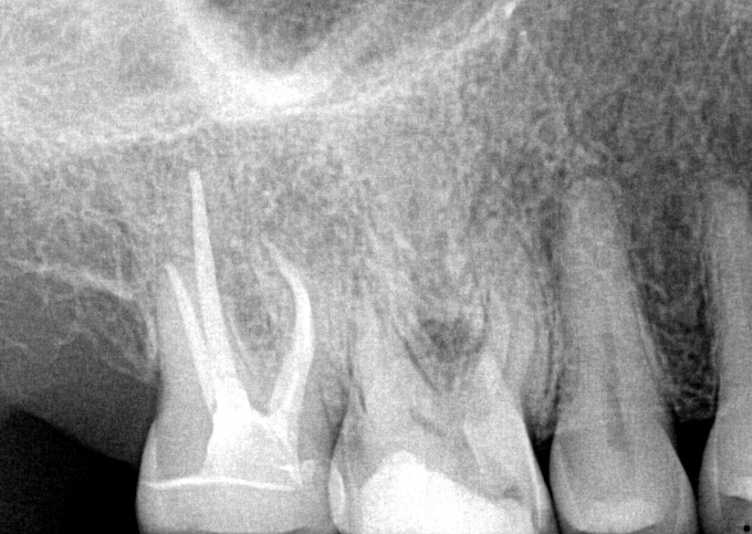
 I'm thinking that there must be internal resorbtion and that this tooth may need extraction and a graft for a future implant. I go and take a scan on him. Check out the tangential view on this screen shot (I put a white arrow on where to look):
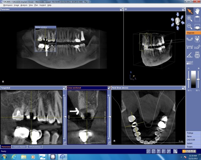
It appears there is a lingual perforation from internal resorbtion. I look at the PA to see how I could overlook something so huge. When I went back, I noticed the darkening in the furca area, but it probed normal and pt never complained of any discomfort so I didn't pay it any mind. If I would have scanned him first, I could have just planned for the extraction and graft instead of wasting all the time I did with the other stuff. Amazing what 3D can show you!!!!